facebook link iconInstagram link iconyoutube channel link iconlinkedin link icon
email and contact iconicon for location
HIP

X-rays, CT Scans and MRIs

by Cory Calendine, MD, Orthopaedic Surgeon, Hip and Knee Specialist

Diagnostic imaging techniques can help delineate musculoskeletal injury and disease. The most commonly used techniques include xrays, computed tomography (CT) scans, and magnetic resonance imaging (MRI).

These imaging tools allow surgeons to "see" inside the body and get a "picture" of the bones, organs, muscles, tendons, nerves, and cartilage.

Xrays

Due to cost and availability, xrays (radiographs) are the most commonly ordered diagnostic imaging modality. Even if a more complex test is required for final diagnosis, xray is often used as a first-line screen.

Xrays use low doses of radiation to image dense objects including bones, calcifications, some tumors, and other dense matter. More dense objects appear white or lighter on xray films  because they absorb the radiation. Less dense soft tissues structures allow the radiation pass-through, therefore appear darker on xray films.

Computed tomography (CT)

CT is an imaging technique that combines xray images from various angles with computer technology that produces more detailed, cross-sectional images of the body. CT scans allow surgeons to visualize 3D size, shape, and position of structures that are deeper within the body and joints. CT scans are more expensive and take more time than regular xray imaging.

Magnetic Resonance Imaging (MRI)

Magnetic resonance imaging (MRI) uses radio waves to produce detailed cross-sectional images of the body. Unlike xrays and CT scans, MRI does not require the use of  radiation. MRI machines use magnetic fields and computer processing to take high-resolution pictures of bones and soft tissues.

Due to the ability to generate more detailed images – especially of soft tissues - MRI can be used to better evaluate ligaments, tendons and cartilage surfaces. MRI scans typically take longer and are more expensive than xrays and CT scans.
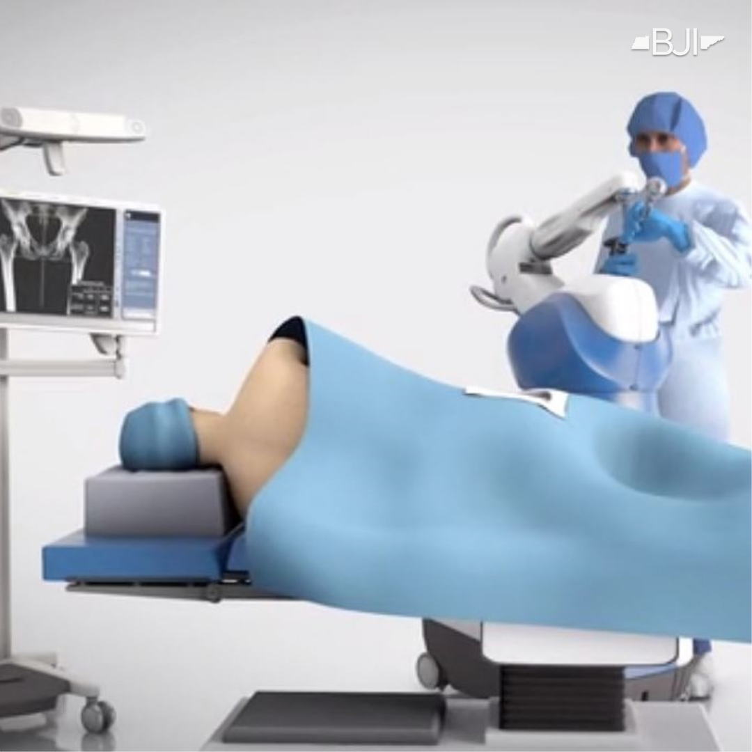
3D Modeling

Increasingly 3D modeling is used to provide surgeons with a precise 3D image of bone and joint structures. 3D modeling is accomplished with sophisticated computer programs and based on actual patient imaging (xray, CT, and/or MRI). As an example, we often use computer-generated 3D modeling (based on CT scans) to complete preoperative planning for knee and hip replacement surgeries.

Thank You for Subscribing to Bone Health & Harmony Blog!
Oops! Something Went Wrong, Please Enter Your Email Again.
Cory Calendine, MD is an Orthopaedic Surgeon and founding partner of the Bone and Joint Institute of Tennessee at Williamson County Hospital in Franklin, TN. Dr. Calendine is an expert in Joint Replacement, specializing in Hip and Knee Surgery. From diagnosis through treatment, the Orthopaedic Surgical experts at the Bone and Joint Institute use the latest techniques and technology to improve care for people with musculoskeletal problems. For more information, please contact our office or schedule your appointment today.  
HIP

Robotic-Assisted Hip Replacement Advantages

If you’re one of the millions of Americans suffering from pain caused by joint arthritis, injury and pain, Mako Robotic-Arm Assisted Surgery might be right for you. Dr. Cory Calendine, Orthopaedic Surgeon, and the team at the Bone and Joint Institute of Tennessee offer state-of-the-art robotic surgery solutions for joint pain sufferers. LEARN MORE.

Read More
HEALTH/WELLNESS

Scorpion Venom: Future Arthritis Treatment?

Scorpion venom could be used to manage arthritis symptoms in the future, according to new research published in the journal Science Translational Medicine. Scientists have successfully used a venom mini-protein with a strong affinity for joint cartilage to deliver anti-inflammatory medication directly into arthritic joints while minimizing side effects in other body tissues.

Read More
KNEE

PRP Injections for Knee Arthritis

PRP injections for knee arthritis offer a promising alternative for patients seeking relief without surgery, using your body's own healing mechanisms to reduce pain and inflammation. This comprehensive guide explains how platelet-rich plasma therapy works, what to expect during treatment, and why many patients experience 6-12 months of improved function and pain relief. Learn about the science behind PRP, success rates showing 60-70% of patients benefit, how it compares to steroid injections and other treatments, and whether you're a good candidate for this innovative approach. With proper recovery support and realistic expectations, PRP therapy can help you return to activities you love while potentially delaying or avoiding knee replacement surgery.

Read More